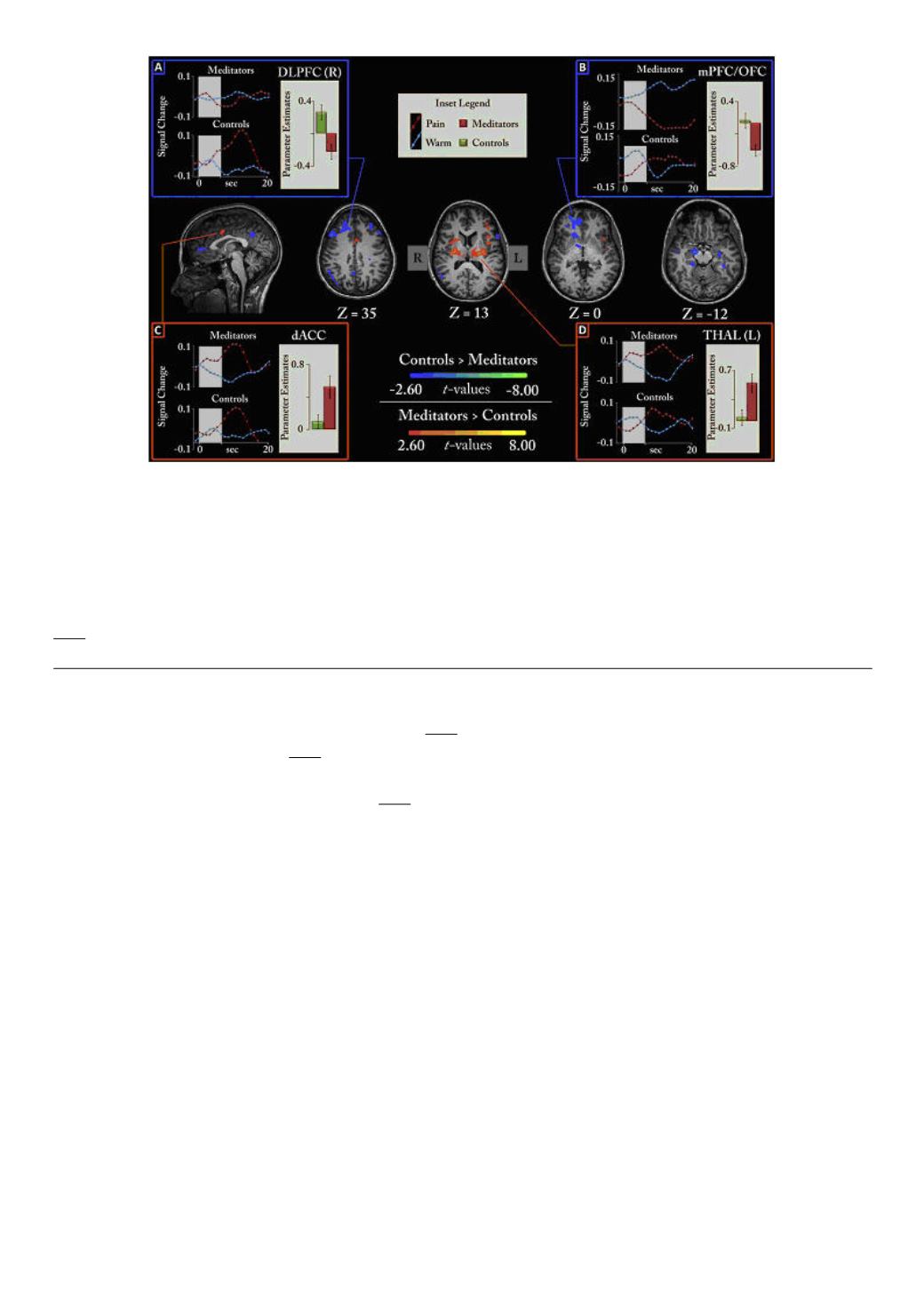
Figure 1.
Differences between meditators and non-meditators during pain in a non-meditative state. Central MRI
images show brain regions statistically different during pain (orange–yellow: meditators > controls), (blue–green:
controls > meditators). Corner insets show average activity levels for each group (right side of each box) as well as the
time course of the activity (left side) during pain trials (red) and warm trials (blue) for each group separately. Gray bars
represent where the stimulus was presented. A: right DLPFC, B: right med-PFC/OFC, C: left THAL, D: dACC spanning
the midline. R = right, L = left.
Taken from Grant J.A., Courtemanche J., Rainville P. A non-elaborative mental stance and decoupling of executive and
pain-related cortices predicts low pain sensitivity in Zen meditators.
2011 Jan;152(1):150-6. This figure has been reproduced with permission of the International Association for the
Study of Pain (IASP). The figure may not be reproduced for any other purpose without permission.
The orbitofrontal cortex is known to receive sensory input (from all modalities) and integrate the
relative value or importance for the individual
The amygdala is a key player in processing
emotion, most famously fear
The hippocampus is a memory-related structure and works in
concert with the dorsolateral-prefrontal cortex, whereas the medial-prefrontal cortex has been
implicated in self-referential processing
So what might all this mean?
While many potential explanations certainly exist for these results, the pattern of brain activity
maps extremely well onto the concept of mindfulness. First, mindfulness should involve monitoring
of the present experience (in this case pain), which would result in activation of the relevant
cortices (ACC, INS, SI, etc.). Second, monitoring occurs in the present moment and likely
precludes the production of elaborate mental narratives. Thus, one could postulate that this mental
stance would not require much in terms of memory or self-related processing, possibly accounting
for the hippocampal, dorsolateral- and medial-prefrontal activity reductions. A reduction in memory
processing should also limit stimulus evaluation, which necessarily involves comparison of present
to past or future. This may explain the orbitofrontal reduction. Finally, if there is less, or no,
evaluation and/or elaboration of the stimuli, one may be less likely to have a strong emotional
reaction, which could explain the amygdala reduction. While this would be a fitting explanation,
given the group being investigated, it is admittedly speculative and will require verification in future
studies with some kind of performance-based measures.
One final result from that study is of particular interest. We examined whether the brain areas listed
above were active (or inactive) in synchrony with each other during pain and whether this differed
between groups. We discovered that, during pain, meditators “functionally” disconnected the PFC
(dorsolateral) with a central pain-related region, the ACC. Information is thought to flow between
260


